Recent advances in electron cryomicroscopy (cryo-EM) have made it possible to visualize extremely complex biological systems, providing novel molecular insight into human biology and disease. Cryo-EM has become an essential structural biology tool, enabling the determination of 3D structures for large biological macromolecules, complexes, and cellular components. At CSHL, the Cyro-EM Facility offers researchers access to cutting-edge instruments that can be applied broadly to study macromolecular machines.
Explore all the CSHL Core Facilities
Core Facility Staff
Facility Head
Facility Manager
Dennis Thomas
thomas@cshl.edu
Equipment
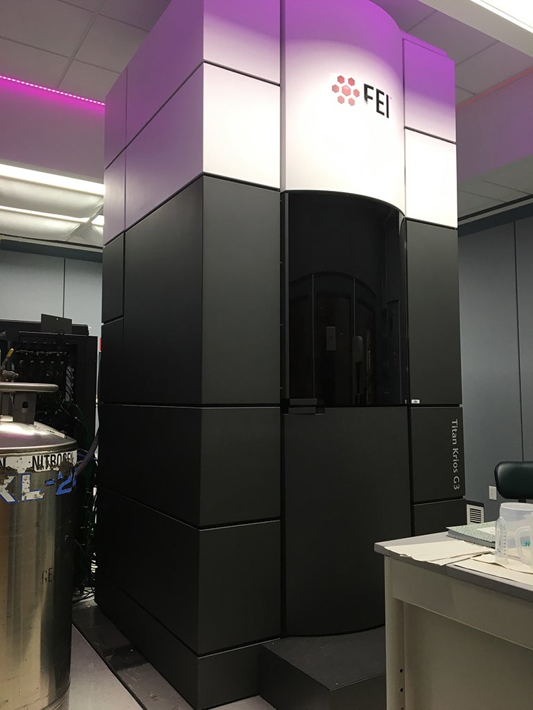
The FEI Titan G3 Krios installation began on August 10th, 2017, in a specially built facility in the Beckman lab. The microscope was accepted early in Dec. 2017, and after training and testing, the cryoEM facility has been operating almost continuously since Jan. 2018. The Titan G3 can collect data automatically. It has the autoloading capability, which can hold and transfer up to 12 samples. To date, we have achieved resolutions as high as 1.8 angstroms and can collect as many as 470 images per hour (350+ typical) using the installed Gatan K3 electron detector. The K3 detector is mounted on a Gatan Bioquantum energy filter. The Titan G3 also has an installed phase plate.
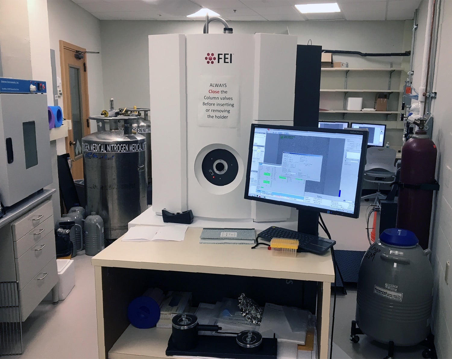
We have a Microscope for screening, an FEI/ThermoFisher Talos 120C for negative stain, and cryo-prescreening of samples.
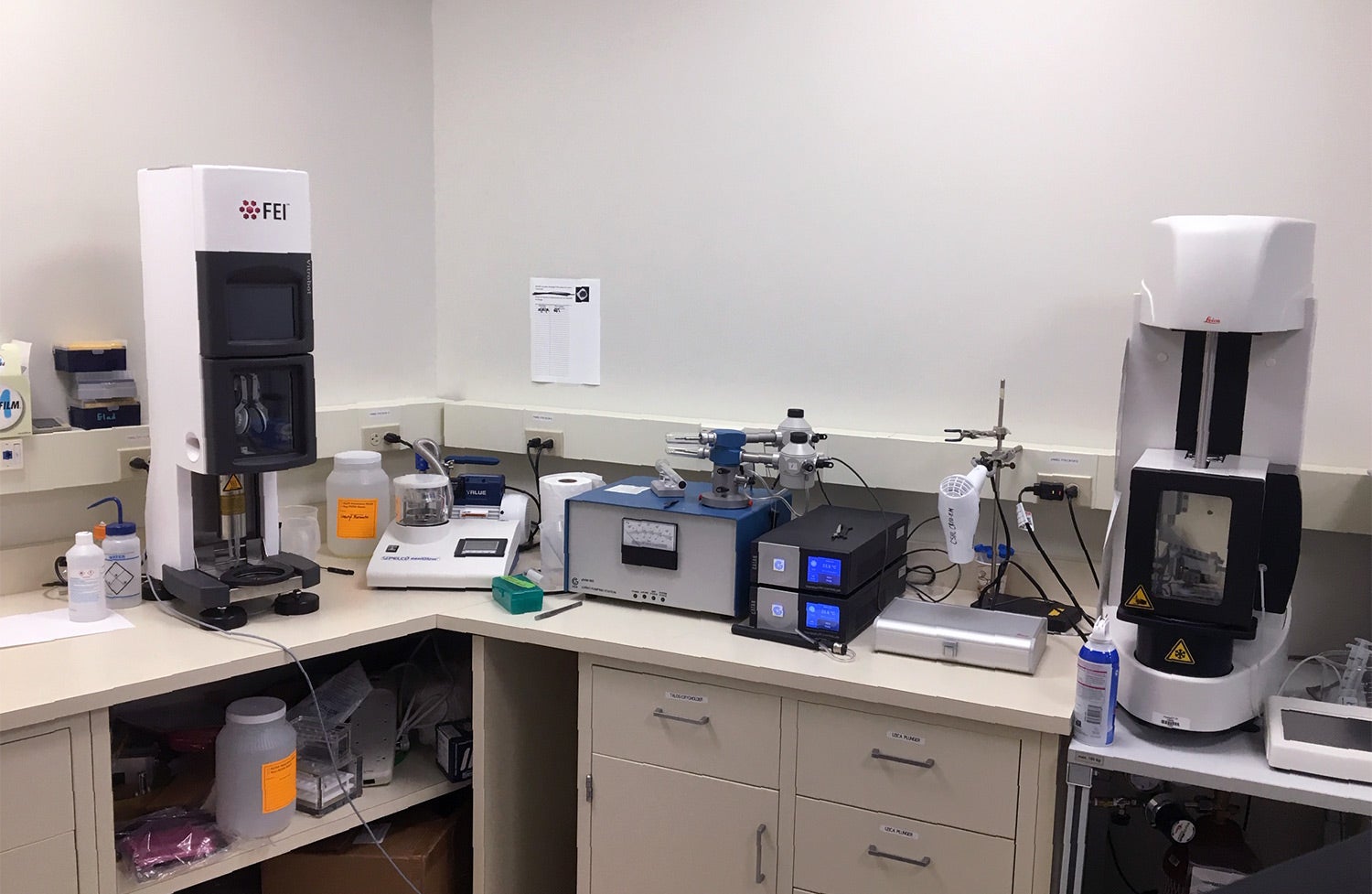
We also have two automated plunge freezers for vitrifying specimens. The FEI/ThermoFisher Vitrobot uses double-sided blotting, and the Leica GP plunger uses single-sided blotting, providing flexibility in sample prep.
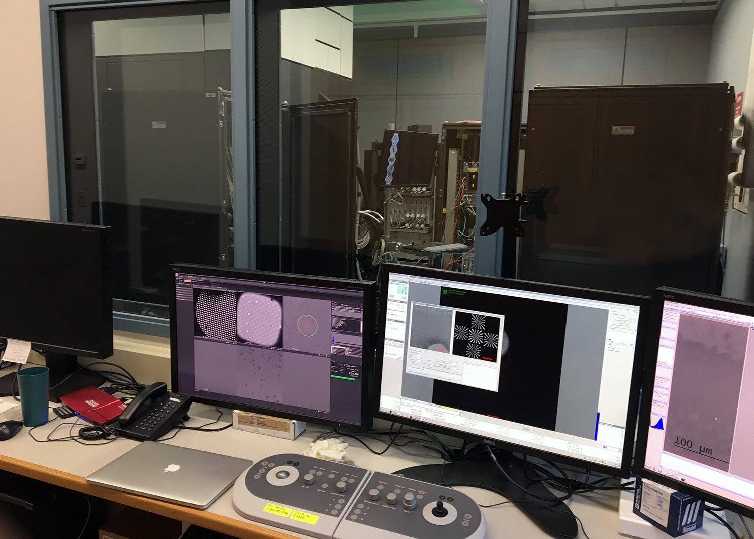
The facility is routinely capable of collecting data at about 350 images per hour or higher, allowing as many as 5000 images to be collected overnight. This means more than 1,000,000 particles are obtained overnight for various samples. The facility has on-the-fly processing capability using a local machine with 4 GPUs and 48 CPUs with 512 GB RAM. This computer is directly connected to the K3 processing computer and then connected via a fiber optic network to our High-performance computing center for access to GPU and CPU clusters, where currently available software is available as part of the SBgrid network.
If you have a project that could benefit from these resources, please contact the Facility Manager, Dennis Thomas, at thomas@cshl.edu or call 516-367-5909. (Ext. 5909)
The Titan G3 operating in Beckmann