Knowing exactly what’s inside a tumor can maximize our ability to fight cancer. But that knowledge doesn’t come easy. Tumors are clusters of constantly changing cancer cells. Some become common cancer variants. Others morph into deadlier, drug-resistant varieties. No one truly understands what governs this chaotic behavior.
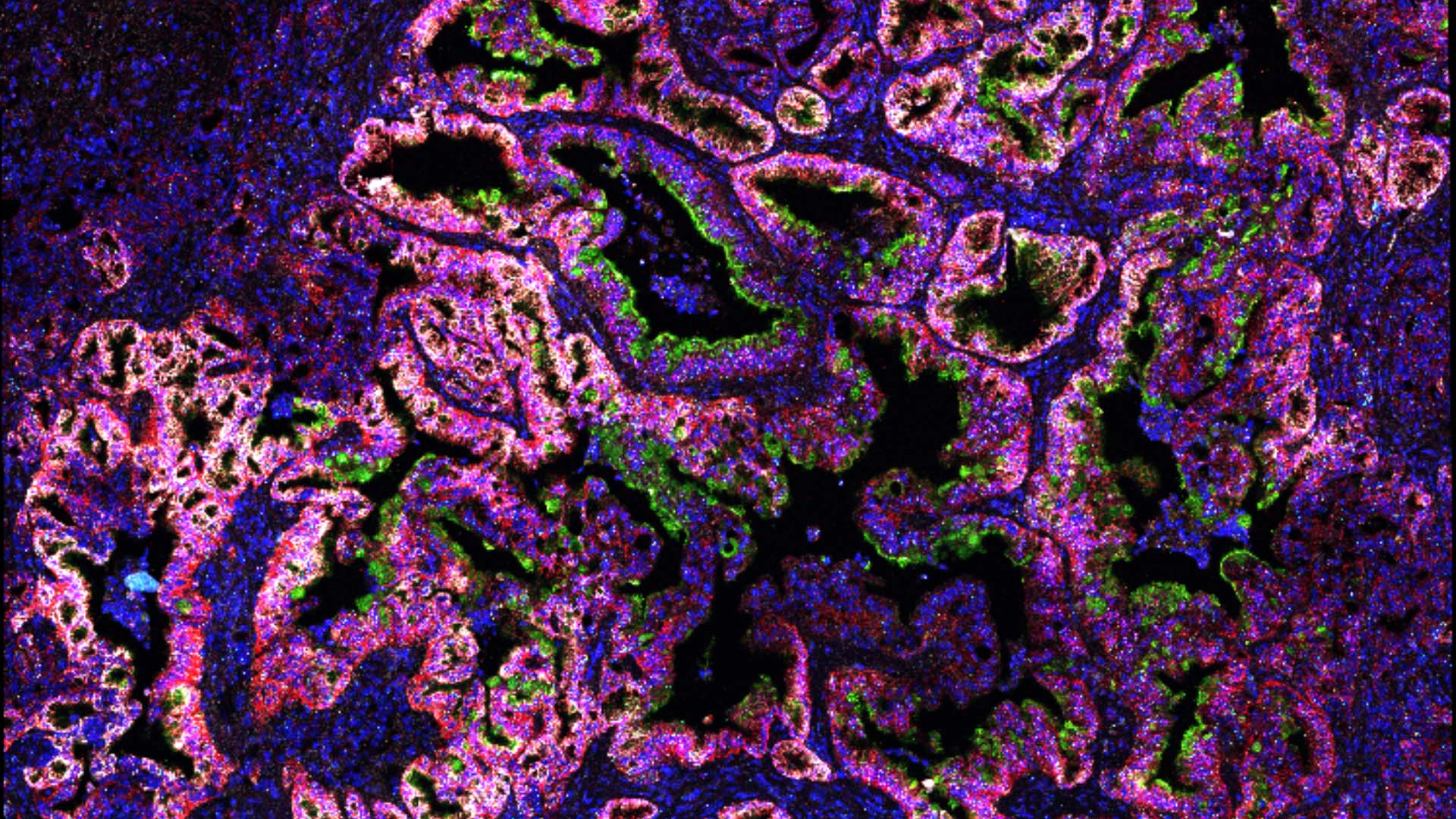
Now, Cold Spring Harbor Laboratory (CSHL) Professor David Tuveson and his team have uncovered a mechanism involved in pancreatic cancer transformation—mucus. During the disease’s early stage, pancreatic cancer cells produce mucus. Additionally, these cells depend on the body’s regulators of mucus production. This new knowledge could help set the stage for future diagnostic or therapeutic strategies.
The unpredictable, shifting nature of tumors makes it challenging to pinpoint the right treatments for patients. “We need to better understand this concept of cell plasticity and design therapy that takes this into consideration,” says Claudia Tonelli, a research investigator in the Tuveson lab, who led the study.
To find out what’s behind tumors’ erratic behavior, Tonelli teamed with Jonathan Preall from CSHL’s Single-Cell Biology Facility. Together, the scientists broke down messy clumps of pancreatic tumors into individual cancer cells. From there, they could examine the unique differences between each pancreatic cancer type. And that’s where mucus’ role in cancer differentiation oozed into view.
Making mucus is messy and challenging for cancer cells. It takes a lot of resources to assemble and export these protein blobs. So why do it in the first place? The scientists discovered that low-grade pancreatic cancer cells of a common variety, known as the classical type, depend on mucus to survive and thrive. As the cancer cells mature and transform into the deadlier type, known as basal-like, they seem to grow out of this dependency. Tonelli suspects mucus may provide fledgling cancer cells with protection against the immune system.
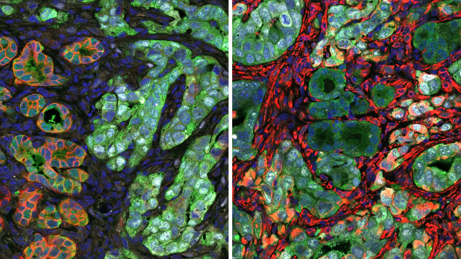
While therapeutically targeting mucus in young, vulnerable pancreatic cancer cells may have some advantages, it’s a double-edged sword. When mucus production is blocked, cancer cells stop growing. But this forces some of them to become the deadlier basal-like cancer cells as a survival mechanism.
“We would have to do further studies to be ready to hit the cancers once they have undergone this differentiation,” says Tonelli. “Identifying a combination of therapies may be an option.”
Knowing what makes pancreatic cancer cells transform may someday help researchers discover better therapeutics. So, while mucus might not be the key to cracking pancreatic cancer, the answer might yet be found right under our nose.
Written by: Luis Sandoval, Communications Specialist | sandova@cshl.edu | 516-367-6826
Funding
Lustgarten Foundation, Thompson Foundation, Pershing Square Foundation, CSHL-Northwell Health Affiliation, Cold Spring Harbor Laboratory Association, National Institutes of Health, Simons Foundation, American-Italian Cancer Foundation, National Cancer Institute
Citation
Tonelli, Claudia., et al., “A mucus production programme promotes classical pancreatic ductal adenocarcinoma”, Gut, Jan 23, 2024. DOI: 10.1136/gutjnl-2023-329839
Core Facilites
Principal Investigator

David Tuveson
Professor
Roy J. Zuckerberg Professor of Cancer Research
Cancer Center Director
M.D., Ph.D., Johns Hopkins University, 1994