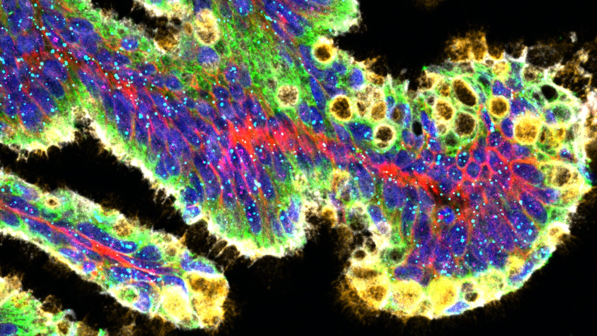
Is this a scene straight out of the latest Star Trek? No, think much more down-to-earth. What we’re seeing here isn’t the Starship Enterprise on fire in the void. It’s a cross-section of precancerous lesions in the pancreas. These mutated cells have a higher risk of turning into cancer than their healthy counterparts. The image above comes to us courtesy of Cold Spring Harbor Laboratory’s (CSHL’s) Tuveson lab and Microscopy Core Facility.
Like the intrepid explorers in Starfleet, CSHL Professor David Tuveson and his team are pushing the frontiers of pancreatic cancer research. In 2015, the Tuveson lab pioneered the study of pancreatic cancer organoids—tiny lab-grown tumors. This led to the first mouse models for two types of the deadly disease. These innovations have since become invaluable in preclinical pancreatic cancer research. Recently, the team made another fascinating discovery. They found that mucus plays a key role in the progression of certain types of pancreatic tumors. Yes, that mucus.
“Early-stage pancreatic cancer cells regulate mucus production essentially the same way as cells in your nose, eyes, lungs, and even your intestines,” says Claudia Tonelli, a research investigator in the Tuveson lab.
To get a closer look at how these mutated cells use and produce mucus, the Tuveson lab partnered with Jonathan Preall from CSHL’s Single-Cell Biology Facility. They found that regulating mucus production is essential for the growth and survival of certain types of pancreatic cancer. But the discovery is a double-edged sword. Shutting off mucus regulation slows tumor growth, but also drives cancer cells to transform into deadlier, drug-resistant varieties.
Single-cell sequencing and microscopy aren’t going to cure cancer on their own. Yet, they do provide unprecedented insight into the disease. These technologies make it possible to pinpoint cancer-related proteins (above, in red, green, and white), mucin (the stuff mucus is made of, in yellow), and even individual mucus-related RNA (the cyan dots), with striking clarity. And that empowers Tuveson and his team at CSHL to go where no cancer researchers have gone before.
Written by: Nick Wurm, Communications Specialist | wurm@cshl.edu | 516-367-5940
About

David Tuveson
Professor
Roy J. Zuckerberg Professor of Cancer Research
Cancer Center Director
M.D., Ph.D., Johns Hopkins University, 1994