Pancreatic cancer has a lot of nerve. Notoriously tricky to detect, the disease also often resists traditional therapy. So, researchers are urgently looking for new ways to disrupt tumor formation. Though scientists know that the nervous system can help cancer spread, its role in the disease’s earliest stages remains unclear. “One phenomenon that is known is called perineural invasion,” says Jeremy Nigri, a postdoc in Professor David Tuveson’s lab at Cold Spring Harbor Laboratory (CSHL). “This means cancer cells will migrate within the nerve and use the nerve as a way to metastasize.”
Now, Nigri and his colleagues at CSHL have discovered that the nervous system plays an active part in pancreatic cancer development, even before tumors form. Using 3D imaging, they found that tumor-promoting fibroblasts called myCAFs send out signals to attract nerve fibers. The myCAFs and nerve cells then work together within pancreatic lesions to create a favorable environment for cancer to grow. The findings are reported in Cancer Discovery, a journal of the American Association for Cancer Research.
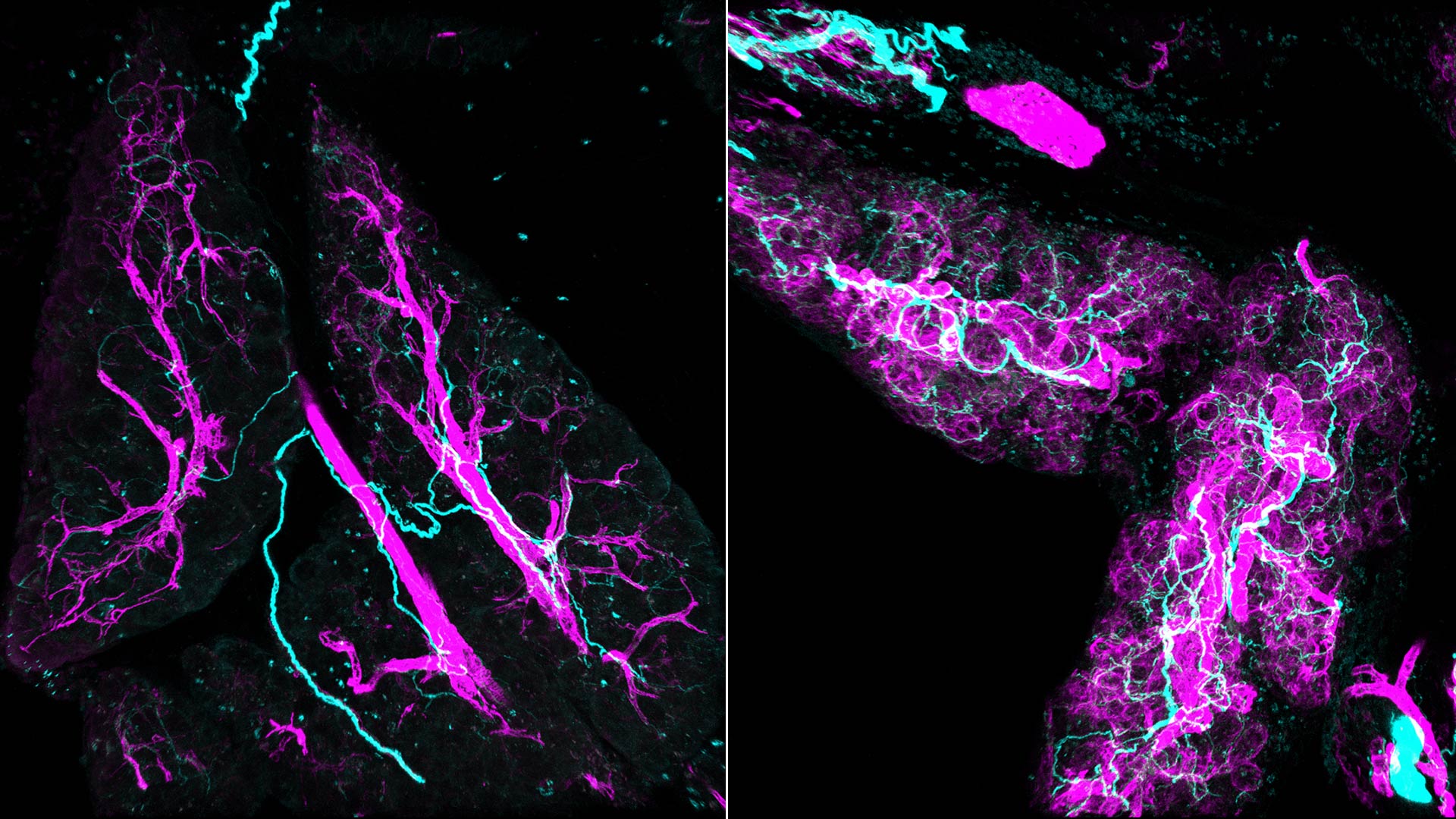
A technique called whole-mount immunofluorescence enabled Tuveson’s team to take 3D photographs of the lesions and surrounding cells. Where standard 2D images show thin nerve fibers as scattered tiny dots, the 3D images reveal a dense network of nerves snaking through and around the myCAFs and lesions. “When we first saw this picture, I was shocked,” Nigri says. “I couldn’t even imagine the lesion like this. I’d only ever seen it in 2D.”
3D images reveal the highly innervated microenvironment of pre-cancerous pancreatic lesions, seen here as red bubbles. Note the network of nerves in cyan and myCAFs in white.
Nigri and his colleagues ran a series of experiments on mice and human cells that uncovered a vicious cycle between myCAFs and nerves. They found myCAFs give off signals that attract nerve fibers from the sympathetic nervous system, which is responsible for our fight-or-flight response. These nerve fibers release the neurotransmitter norepinephrine, which binds to the fibroblasts and triggers a calcium spike that further activates myCAFs. This spike not only promotes pre-cancerous growth, but also pulls in even more nerve fibers, locking the system into a dangerous self-reinforcing loop.
“In one experiment, we use a neurotoxin to disable the sympathetic nervous system,” Nigri says. “We show reduced fibroblast activation and a nearly 50% reduction in tumor growth.”
Because the myCAF-nerve loop happens so early, disrupting this cycle could lead to potential new therapies. The findings suggest that clinically available drugs, including doxazosin, may be effective when combined with standard treatments like chemotherapy or immunotherapy. “The next step will be to study this more in detail and try to find a way to block the crosstalk between fibroblasts and nerves,” Nigri says. “With support from groups like the Lustgarten Foundation and Pancreatic Cancer Action Network, we hope to one day help improve patient outcomes.”
Written by: Margaret Osborne, Science Writer | publicaffairs@cshl.edu | 516-367-8455
Funding
National Institutes of Health, Defense Health Agency, Lustgarten Foundation, Thompson Foundation, Pershing Square Foundation, Simons Foundation, CSHL-Northwell Health Affiliation, Cold Spring Harbor Laboratory Association, U.S. Department of Defense, Pancreatic Cancer Action Network, National Cancer Institute, Donaldson Foundation, Northwell Health Cancer Institute
Citation
Nigri, J., et al., “Myofibroblasts induce neuroplasticity to promote pancreatic inflammation and cancer progression”, Cancer Discovery, February 9, 2026. DOI: 10.1158/2159-8290.CD-25-1337
Core Facilites
Principal Investigator

David Tuveson
Professor
Roy J. Zuckerberg Professor of Cancer Research
Cancer Center Director
M.D., Ph.D., Johns Hopkins University, 1994