Branching isn’t just for trees. This biological process occurs in animal development, enabling organs to perform complex functions. Branch-like structures form in lungs, kidneys, and breasts, among other places. Importantly, only in female mammary glands does most branching occur years after birth. It happens during puberty and again during pregnancy as milk ducts branch out in preparation for breastfeeding. Disturbances here have been linked to breast cancer. However, studying branching can be difficult and time-consuming.
Now, Cold Spring Harbor Laboratory (CSHL) researchers have developed a tool to quickly quantify changes in the branches of mouse mammary glands. The system, called MaGNet, was created by Steven Lewis, Lucia Téllez Pérez, and Samantha Henry, three graduate students in CSHL’s dos Santos lab. It could one day be used to study how hormonal changes and treatments affect mammary glands or even to detect early warning signs of breast cancer.
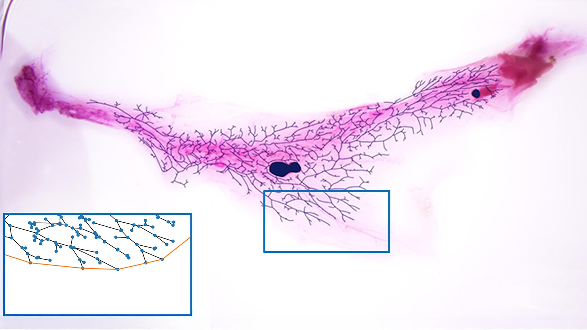
Lewis had the idea for MaGNet—the Mammary Gland Network analysis tool—after seeing CSHL Associate Professor Saket Navlakha give a talk at the Lab. Lewis wondered if a mathematical model that Navlakha’s team applied to plants could be translated to mammary glands. “It felt like a natural connection to another branching structure,” he says.
Typically, researchers studying mouse mammary glands need to cut thin slices of breast tissue, analyze them under a microscope, and manually count the ducts and branches in each slice. The process is “very time-consuming and sometimes inconsistent,” Henry says. “Often, you don’t get the entire architecture of the mammary gland.”
MaGNet was built to precisely compare stained images of the mammary gland. Researchers working with the system simply trace the branches and use software called NetworkX to plot them as networks. From there, computer code analyzes the networks and quantifies the data. “With this tool, we can measure the total length of the ductile tree as well as the number of ducts, alveoli, and branching structures,” Téllez Pérez says. “It’s very easy to quickly plot different networks and run tests.”
Right now, MaGNet is only used in mice. However, the code could be tweaked for any branching system. The researchers envision MaGNet eventually being used to learn how certain conditions, like infections—or life events, like pregnancy and menopause—affect cancer risk. It could also help with early diagnosis. “We’re always hunting for warning signs that arise before you can feel a lump or see anything on a mammogram or ultrasound,” Lewis says. “Imagine an automated tool could say there’s no tumor yet, but there are changes detectable. That’s our hope, our dream.”
Written by: Margaret Osborne, Science Writer | publicaffairs@cshl.edu | 516-367-8455
Funding
Pershing Square Foundation, Simons Foundation, CSHL-Northwell Health Affiliation, National Institutes of Health, National Cancer Institute, “la Caixa” Foundation
Citation
Lewis, S., et al., “MaGNet: A Network-Based Method for Quantitative Analysis of the Mammary Ductal Tree in Developing Female Mice”, Journal of Mammary Gland Biology and Neoplasia, September 26, 2025. DOI: 10.1007/s10911-025-09589-1
Core Facilites
Principal Investigator

Camila dos Santos
Associate Professor
Cancer Center Program Co-Leader
Ph.D., Universidade Estadual de Campinas - Brazil, 2007