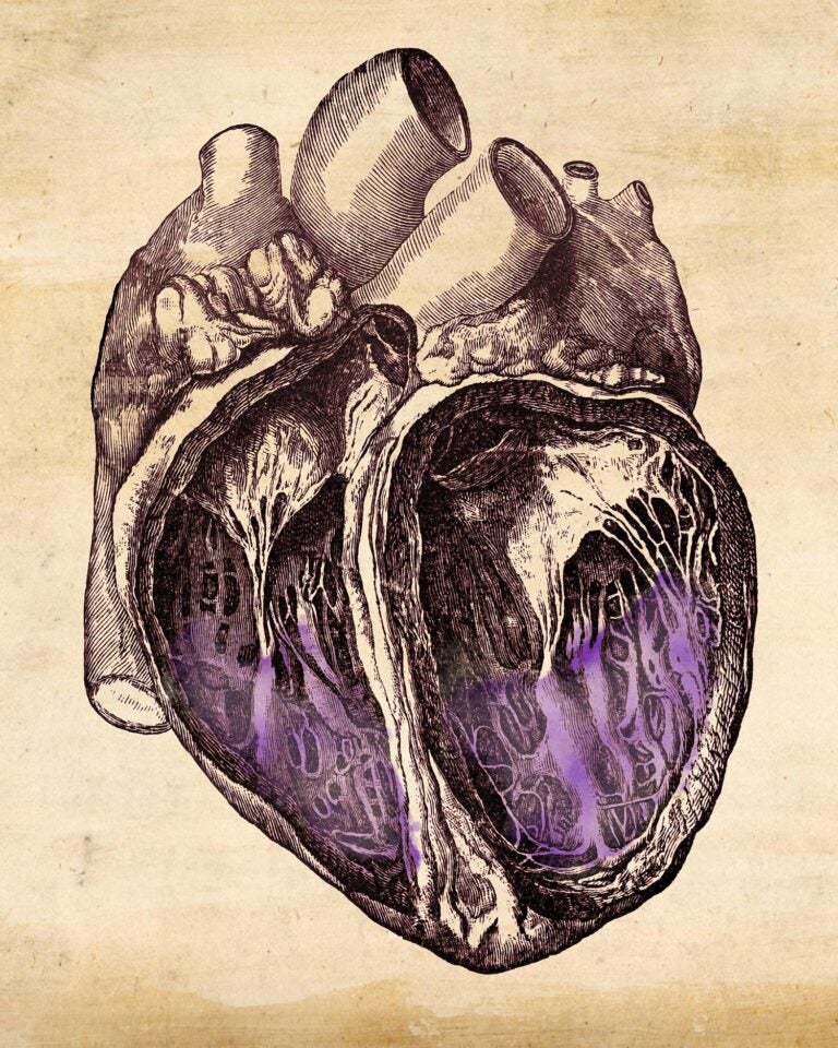
Researchers have investigated the function of a complex mesh of muscle fibers that line the inner surface of the heart. The study, published in the journal Nature, sheds light on questions asked by Leonardo da Vinci 500 years ago, and shows how the shape of these muscles impacts heart performance and heart failure.
In humans, the heart is the first functional organ to develop and starts beating spontaneously only four weeks after conception. Early in development, the heart grows an intricate network of muscle fibers—called trabeculae—that form geometric patterns on the heart’s inner surface. These are thought to help oxygenate the developing heart, but their function in adults has remained an unsolved puzzle since the 16th century.

To understand the roles and development of trabeculae, an international team of researchers used artificial intelligence to analyse 25,000 magnetic resonance imaging (MRI) scans of the heart, along with associated heart morphology and genetic data. The study reveals how trabeculae work and develop, and how their shape can influence heart disease. UK Biobank has made the study data openly available.
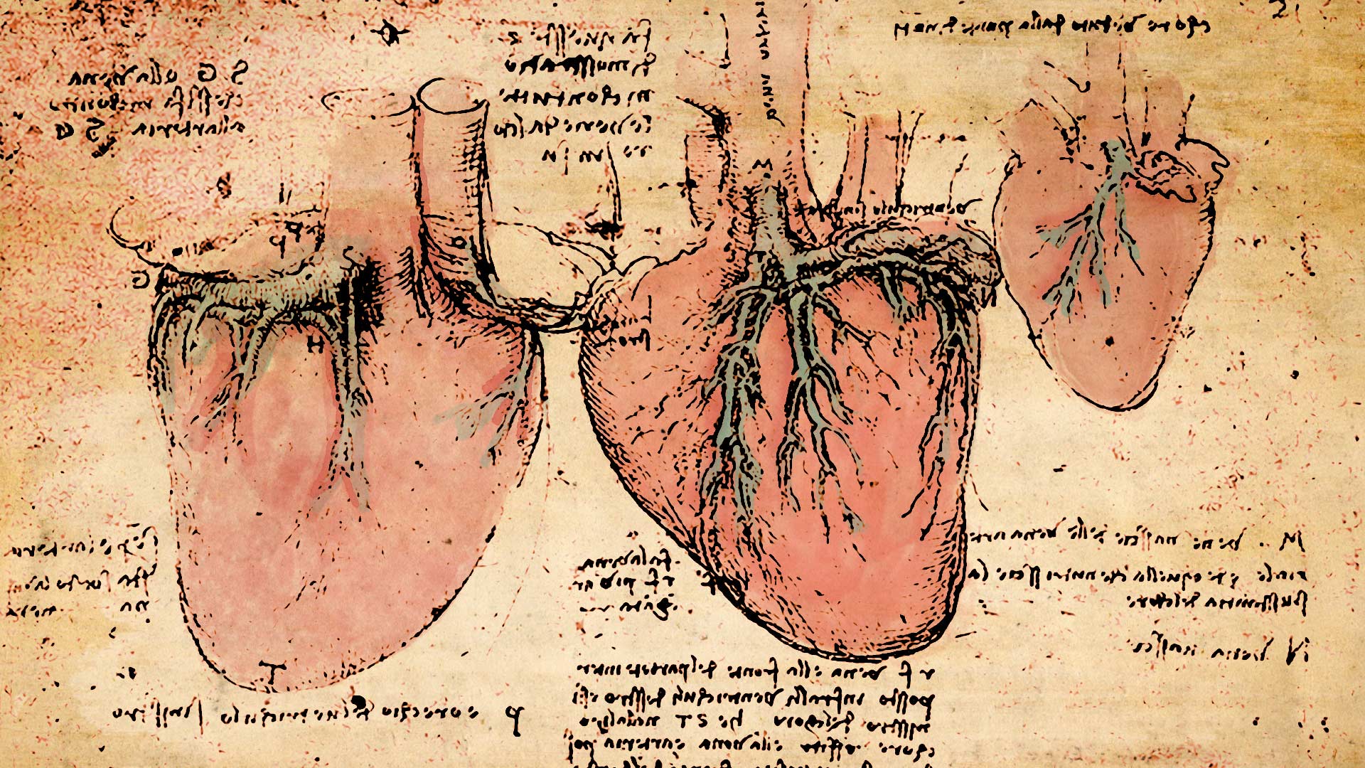
Leonardo da Vinci was the first to sketch trabeculae and their snowflake-like fractal patterns in the 16th century. He speculated that they warm the blood as it flows through the heart, but their true importance has not been recognized until now.
“Our findings answer very old questions in basic human biology. As large-scale genetic analyses and artificial intelligence progress, we’re rebooting our understanding of physiology to an unprecedented scale,” says Ewan Birney, deputy director general of EMBL.

The study also highlights six regions in human DNA that affect how the fractal patterns in these muscle fibers develop. Intriguingly, the researchers found that two of these regions also regulate branching of nerve cells, suggesting a similar mechanism may be at work in the developing brain.
The researchers discovered that the shape of trabeculae affects the performance of the heart, suggesting a potential link to heart disease. To confirm this, they analyzed genetic data from 50,000 patients and found that different fractal patterns in these muscle fibers affected the risk of developing heart failure. Nearly 5 million Americans suffer from congestive heart failure.
Further research on trabeculae may help scientists better understand how common heart diseases develop and explore new approaches to treatment.
“Leonardo da Vinci sketched these intricate muscles inside the heart 500 years ago, and it’s only now that we’re beginning to understand how important they are to human health. This work offers an exciting new direction for research into heart failure,” says Declan O’Regan, clinical scientist and consultant radiologist at the MRC London Institute of Medical Sciences. This project included collaborators at Cold Spring Harbor Laboratory, EMBL’s European Bioinformatics Institute (EMBL-EBI), the MRC London Institute of Medical Sciences, Heidelberg University, and the Politecnico di Milano.
This story originally appeared in the Winter 2020 edition of the Harbor Transcript magazine.
Funding
RCUK/Medical Research Council (MRC); British Heart Foundation (BHF); Wellcome Trust; National Institute of Environmental Health Sciences; Heidelberg University; Simons Center for Quantitative Biology at Cold Spring Harbor Laboratory; National Institute for Health Research Biomedical Research Centre; Edmond J. Safra Foundation and Lily Safra; National Institute for Health Research; UK Dementia Research Institute, UK Research and Innovation Health; Research Center for Molecular Medicine (HRCMM); Deutsche Herzstiftung e.V.; and Joachim Herz Stiftung.
Citation
Meyer, H. et. al., “Genetic and functional insights into the fractal structure of the heart”, Nature, August 19, 2020. DOI: 10.1038/s41586-020-2635-8

