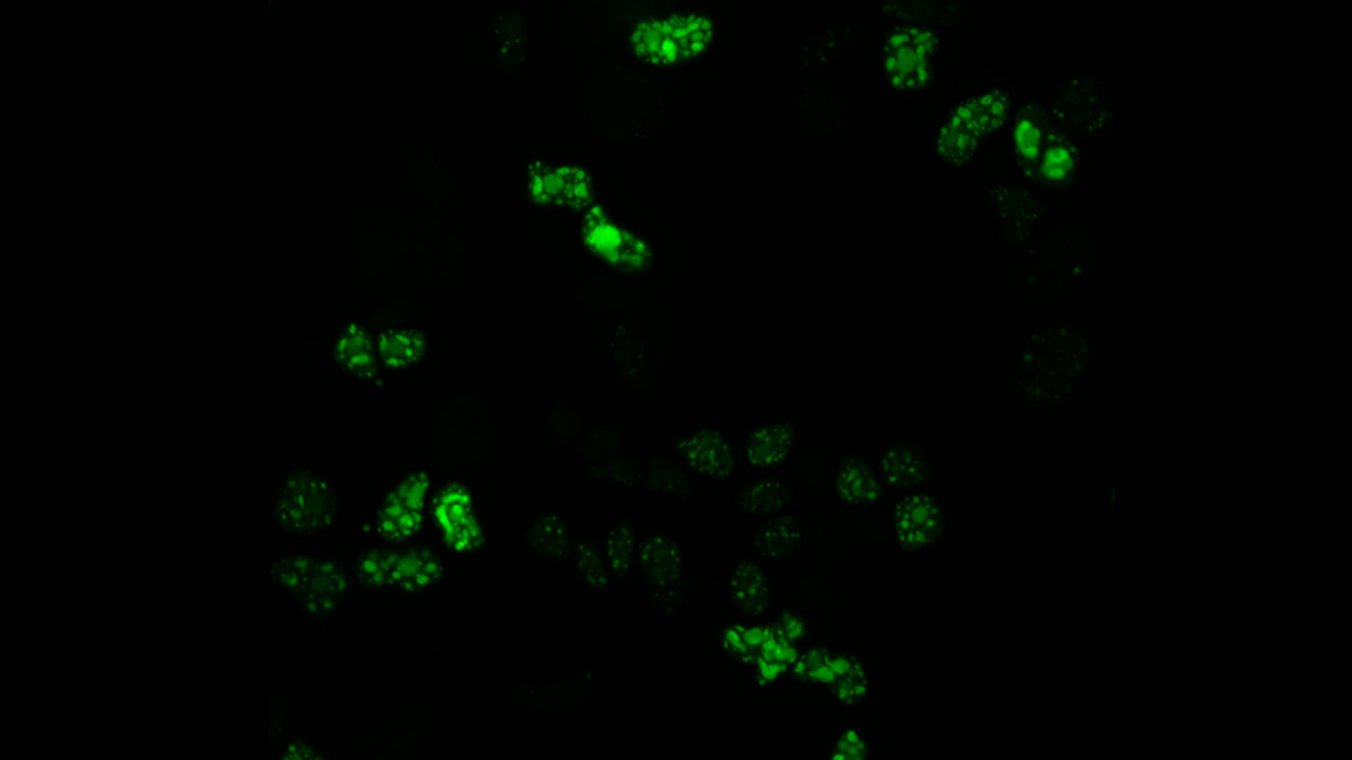
Bodybuilders and cellular mechanisms agree generating protein is a heavy lift. To complete the task, cells rely on complexes called spliceosomes. These molecular machines snip extra bits out of our genes’ RNA copies and piece together precise instructions for protein-building. When the splicing process goes awry, it can result in diseases like cancer or spinal muscular atrophy. Cold Spring Harbor Laboratory (CSHL) Professor Adrian Krainer helped develop the first FDA-approved treatment for this devastating genetic disorder. Now, his team has discovered that two important regulator proteins work together to keep the splicing process on track.
One of those regulators, a protein called SRSF1, has been a focus of Krainer’s lab since 1990. It was then that he discovered cells need SRSF1 for splicing to occur. His team has since found that too much SRSF1 can prompt cells to grow cancerous. Given this link to human disease, Krainer has been trying to determine just how SRSF1 works.
In his latest study, Krainer and graduate student Danilo Segovia set out to identify proteins that commingle with SRSF1 inside cells. They turned to a new technique that lets researchers track any protein that comes close to their molecule of interest—no matter how briefly. That’s important, Segovia says, because splicing is a dynamic process. Many molecules come and go quickly as the spliceosome assembles and does its work. With the new method, “You get the history of all the proteins that were within close range of SRSF1,” Segovia says.
RNA splicing might seem difficult to comprehend. However, when something goes wrong with it, the impacts can be clear as day, severely impacting people’s lives. Today, CSHL scientists are trying to reach a better understanding of the proteins that regulate this complicated process.
‘History’ is the right word, as the list turned out quite long. It included known splicing regulators as well as some surprises. Segovia and Krainer were particularly interested in SRSF1’s interaction with a protein called DDX23. This enzyme helps newly built spliceosomes get themselves into shape for RNA splicing.
Next, Krainer and Segovia teamed with CSHL Director of Research Leemor Joshua-Tor. Together, they confirmed a strong interaction between SRSF1 and DDX23. Segovia proposes that their connection might be important for ensuring DDX23 is in the right place at the right time or in the right form to promote splicing.
“SRSF1 seems to be pretty central here,” Krainer says. He explains that in addition to its interaction with DDX23, the regulator protein is needed for an earlier step in spliceosome assembly. “It seems to be at this pivotal point between spliceosome transitions.” With his group’s list of SRSF1-interacting proteins, researchers now have a new set of clues into how this critical regulator does its work. That could help reduce the lift that comes with looking for new insights into cancer and other devastating diseases.
Written by: Jennifer Michalowski, Science Writer | publicaffairs@cshl.edu | 516-367-8455
Funding
National Institutes of Health, National Cancer Institute
Citation
Segovia, D., et al., “SRSF1 interactome determined by proximity labeling reveals direct interaction with spliceosomal RNA helicase DDX23”, PNAS, May 14, 2024. DOI: 10.1073/pnas.2322974121
Core Facilites
Principal Investigator

Adrian R. Krainer
Professor
St. Giles Foundation Professor
Cancer Center Program Co-Leader
Ph.D., Harvard University, 1986
