Diagnostic Applications Foreseen in Cancer, Neurological and Psychiatric Illnesses
Cold Spring Harbor, NY — A team of scientists including researchers at Cold Spring Harbor Laboratory (CSHL) have identified and validated the first biomarker that permits neural stem and progenitor cells (NPCs) to be tracked, non-invasively, in the brains of living human subjects. This important advance could lead to significantly better diagnosis and monitoring of brain tumors and a range of serious neurological and psychiatric disorders.
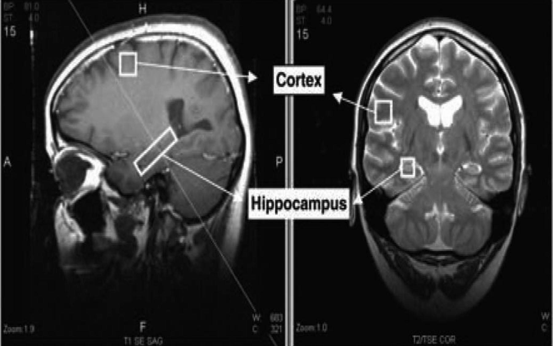
The biomarker is a lipid molecule whose presence the scientists were able consistently to detect in a part of the brain called the hippocampus where new nerve cells are known to be generated. The marker was not detected in the cortex and other parts of the brain where this process, called neurogenesis, does not occur in healthy adults.
As elsewhere in the body, the rise of new cells in the brain is a process that can be traced to stem cells, which, through mechanisms still only partly grasped, give birth to “daughter” progenitor cells that undergo repeated division and maturation into “adult” cells. As recently as a few years ago, most scientists did not believe that new nerve cells were created anywhere in the adult brain.
The newly discovered marker can be detected when NPCs—stem-like “progenitor” cells—are actively dividing, a mark that new nerve cells are being created. “Until now, there was no way to identify and track these cells in living people, to get a dynamic picture of neurogenesis,” said Grigori Enikolopov, Ph.D.
A fuller understanding of neural stem and progenitor cells could one day unlock the secret to nervous-system regeneration following a stroke or massive trauma. In the nearer-term, discovery of the neural stem-cell biomarker just reported is likely to yield more powerful diagnostics.
“The technique the team has developed is based on MRI technology that is currently in widespread use to perform non-invasive scans of the living brain and can tell us where stem-like cells are dividing,” said Dr. Enikolopov, whose CSHL lab specializes in the study of stem cells, in the brain and in other tissues. “Although we are only just beginning to test applications, it is clear that this biomarker may have promise in identifying cell proliferation in the brain, which can be a sign of cancer. In other patients, it could show us how neurogenesis is related to the course of diseases such as depression, bipolar disorder, Alzheimer’s, Parkinson’s, MS, and post-traumatic stress disorder.”
In 2006, Dr. Enikolopov demonstrated that the antidepressant fluoxetine (Prozac) stimulates the creation of new nerve cells in the hippocampus of depressed patients. He later demonstrated that an even more pronounced effect was brought about by other depression treatments, electroconvulsive therapy and deep-brain stimulation.
“The recent finding that neural progenitor cells exist in adult human brain has opened a whole new field in neuroscience,” said Walter J. Koroshetz, M.D., deputy director of the NIH’s National Institute of Neurological Disorders and Stroke (NINDS), which helped fund the work. “The ability to track these cells in living people would be a major breakthrough in understanding brain development in children and continued maturation of the adult brain. It could also be a very useful tool for research aimed at influencing NPCs to restore or maintain brain health.”
Discovery of the neural stem cell marker relied heavily upon the development of an ingenious algorithm devised by Dr. Petar M. Djuric of SUNY Stony Brook. That mathematical formula made the marker’s spectroscopic “image” stand out amid a field filled with visual “noise,” in much the same way as algorithms used in submarine sonar equipment filter out all ambient noise save that of other subs. Filtering out “noise” in the brain enabled the team to demonstrate the presence of the biomarker in live animals and in human subjects.
Written by: Communications Department | publicaffairs@cshl.edu | 516-367-8455
Citation
“Magnetic Resonance Spectroscopy Identifies Neural Progenitor Cells in the Live Human Brain,” appears in Science on November 9. Along with Dr. Enikolopov, Dr. Mirjana Maletic-Savatic of SUNY Stony Brook is a corresponding author of the paper. The compete citation is as follows: Louis N. Manganas, Xueying Zhang, Yao Li, Raphael D. Hazel, S. David Smith, Mark E. Wagshul, Fritz Henn, Helene Benveniste, Petar M. Djuric, Grigori Enikolopov, and Mirjana Maletic-Savatic. The paper is available online at: http://www.eurekalert.org/jrnls/sci/index.php