Cold Spring Harbor, NY — Forget gigabytes. Even the most powerful computers available today are mere playthings compared to the complexity, efficiency, and information processing capacity of the human brain. Underlying the brain’s far superior design are the billion-million or so connections between brain cells—called synapses—that form vast neural networks in which brain cells, or neurons, are each connected to thousands of other neurons. These networks—and their ability to be shaped by experience—enable us to receive, process, store, and retrieve all manner of information about our world. Unfortunately, the extremely tiny size of synapses and the limitations of conventional experimental techniques have hampered detailed studies of these essential structures. (One trillion synaptic compartments, or “dendritic spines,” could fit into a thimble). Now, scientists at Cold Spring Harbor Laboratory have overcome these technical obstacles to gain an extremely close look at the properties of dendritic spines and synapses that govern brain function.
“Our findings reveal fundamental properties of synapses that enables them to trigger the changes in neurons that underlie learning and memory,” says Karel Svoboda, the principal author of the study which will be published tomorrow in Nature. Svoboda, an investigator of the Howard Hughes Medical Institute at Cold Spring Harbor Laboratory, helped pioneer the use of a high-resolution imaging technique called “two-photon microscopy” in neuroscience.
In the current study, Svoboda and his colleague Bernardo Sabatini electrically stimulated brain neurons and used two-photon microscopy to watch as calcium rushed into single dendritic spines of these neurons (see figure). These measurements enabled the researchers to determine the number and type of “calcium channels” present at synapses in a region of the brain important for learning and memory, the hippocampus. Calcium channels are molecular gates that open in response to electrical stimulation and allow calcium to flow into dendritic spines. Calcium, in turn, triggers biochemical events in the spine which modify synaptic strength and thereby encode memories.
In their study, Sabatini and Svoboda could detect if single calcium channels opened or, by chance, remained closed following stimulation. Measuring the probability of channel opening, “like tossing a coin, where heads is open and tails is closed,” says Svoboda, enabled him and Sabatini to determine the number of calcium channels per spine. The scientists discovered that depending on their size, spines contain from one to twenty, and typically three, calcium channels.
“Visually examining calcium fluctuations in a single dendritic spine in the brain as we have done is akin to examining the wrinkles on a raisin sitting on the 50-yard line of a football stadium from the Goodyear blimp,” says Sabatini.
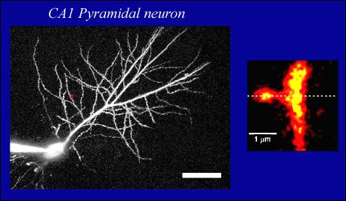
Right: Single dendritic spine, a tiny, bulbous structure that contains a synapse or a connection to another neuron. Yellow blob to the left is a snapshot of calcium rushing into the spine in response to electrical stimulation. This is the basis of brain cell communication, seen here at extremely high magnification
But nothing is that simple in the brain. Which type of channel is causing these changes in calcium? There are at least six known varieties of calcium channels that could be present in spines, each having different properties. Using chemical probes, Sabatini and Svoboda were able to demonstrate that one specific type of channel (the R type) is solely responsible for the influx of calcium that they observed. “We are looking at the behavior of single calcium channels in their natural environment, the brain.” said Sabatini.
“The local influx of calcium we have observed in spines is a fundamental measure of the information carried in one particular brain neuron and how it is processed locally. The information encoded in the messages passed between neurons is simple,” says Sabatini. “It’s not unlike computer programming code where a single command can be either one for a positive response or zero for a negative response.” When an action potential causes a calcium channel to open, that’s a one, when it fails to open that’s a zero.
Scientists believe that the strengthening of synapses between neurons in response to experience ultimately gives rise to networks of neurons that govern complex brain functions like learning and memory. Moreover, communication within these networks forms the basis of thinking and self-awareness that we call cognition. Visualizing how neurons communicate with each other on the most basic level, as Sabatini and Svoboda have done, provides important clues for understanding how our brains outperform the most sophisticated computers and enable us to grasp the human experience.
Written by: Communications Department | publicaffairs@cshl.edu | 516-367-8455